
A novel MRI method developed at MIT permits for detailed imaging of bioluminescence deep throughout the mind, providing new insights into how mind cells develop and talk with one another.
Scientists usually label cells with proteins that glow, permitting them to trace the expansion of a tumor, or measure adjustments in gene expression that happen as cells differentiate.
Whereas this method works properly in cells and a few tissues of the physique, it has been troublesome to use this method to picture buildings deep throughout the mind, as a result of the sunshine scatters an excessive amount of earlier than it may be detected.
MIT engineers have now provide you with a novel technique to detect any such gentle, often called bioluminescence, within the mind: They engineered blood vessels of the mind to precise a protein that causes them to dilate within the presence of sunshine. That dilation can then be noticed with magnetic resonance imaging (MRI), permitting researchers to pinpoint the supply of sunshine.
“A well known drawback that we face in neuroscience, in addition to different fields, is that it’s very troublesome to make use of optical instruments in deep tissue. One of many core targets of our examine was to provide you with a technique to picture bioluminescent molecules in deep tissue with moderately excessive decision,” says Alan Jasanoff, an MIT professor of organic engineering, mind and cognitive sciences, and nuclear science and engineering.
The brand new method developed by Jasanoff and his colleagues might allow researchers to discover the internal workings of the mind in additional element than has beforehand been potential.
Jasanoff, who can be an affiliate investigator at MIT’s McGovern Institute for Mind Analysis, is the senior creator of the examine, which seems right now (Might 10) in Nature Biomedical Engineering. Former MIT postdocs Robert Ohlendorf and Nan Li are the lead authors of the paper.
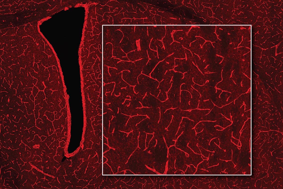
A brand new technique to detect bioluminescence within the mind makes use of magnetic resonance imaging (MRI). The method, developed at MIT, might allow researchers to discover the internal workings of the mind in additional element than beforehand potential. Pictured are blood vessels that now seem shiny pink after transduction with a gene that provides them photosensitivity. Credit score: Courtesy of the researchers
Detecting Mild
Bioluminescent proteins are discovered in lots of organisms, together with jellyfish and fireflies. Scientists use these proteins to label particular proteins or cells, whose glow may be detected by a luminometer. One of many proteins usually used for this goal is luciferase, which is available in a wide range of kinds that glow in numerous colours.
Jasanoff’s lab, which makes a speciality of creating new methods to picture the mind utilizing MRI, needed to discover a technique to detect luciferase deep throughout the mind. To realize that, they got here up with a way for reworking the blood vessels of the mind into gentle detectors. A preferred type of MRI works by imaging adjustments in blood move within the mind, so the researchers engineered the blood vessels themselves to reply to gentle by dilating.
“Blood vessels are a dominant supply of imaging distinction in purposeful MRI and different non-invasive imaging strategies, so we thought we might convert the intrinsic capacity of those strategies to picture blood vessels into a method for imaging gentle, by photosensitizing the blood vessels themselves,” Jasanoff says.
To make the blood vessels delicate to gentle, the researcher engineered them to precise a bacterial protein known as Beggiatoa photoactivated adenylate cyclase (bPAC). When uncovered to gentle, this enzyme produces a molecule known as cAMP, which causes blood vessels to dilate. When blood vessels dilate, it alters the steadiness of oxygenated and deoxygenated hemoglobin, which have completely different magnetic properties. This shift in magnetic properties may be detected by MRI.
BPAC responds particularly to blue gentle, which has a brief wavelength, so it detects gentle generated inside shut vary. The researchers used a viral vector to ship the gene for bPAC particularly to the graceful muscle cells that make up blood vessels. When this vector was injected in rats, blood vessels all through a big space of the mind grew to become light-sensitive.
“Blood vessels type a community within the mind that’s extraordinarily dense. Each cell within the mind is inside a pair dozen microns of a blood vessel,” Jasanoff says. “The way in which I like to explain our method is that we primarily flip the vasculature of the mind right into a three-dimensional digital camera.”
As soon as the blood vessels have been sensitized to gentle, the researchers implanted cells that had been engineered to precise luciferase if a substrate known as CZT is current. Within the rats, the researchers have been in a position to detect luciferase by imaging the mind with MRI, which revealed dilated blood vessels.
Monitoring Adjustments within the Mind
The researchers then examined whether or not their method might detect gentle produced by the mind’s personal cells, in the event that they have been engineered to precise luciferase. They delivered the gene for a sort of luciferase known as GLuc to cells in a deep mind area often called the striatum. When the CZT substrate was injected into the animals, MRI imaging revealed the websites the place gentle had been emitted.
This system, which the researchers dubbed bioluminescence imaging utilizing hemodynamics, or BLUsH, may very well be utilized in a wide range of methods to assist scientists be taught extra in regards to the mind, Jasanoff says.
For one, it may very well be used to map adjustments in gene expression, by linking the expression of luciferase to a selected gene. This might assist researchers observe how gene expression adjustments throughout embryonic growth and cell differentiation, or when new reminiscences type. Luciferase may be used to map anatomical connections between cells or to disclose how cells talk with one another.
The researchers now plan to discover a few of these functions, in addition to adapting the method to be used in mice and different animal fashions.
Reference: “Imaging bioluminescence by detecting localized haemodynamic distinction from photosensitized vasculature” by Robert Ohlendorf, Nan Li, Valerie Doan Phi Van, Miriam Schwalm, Yuting Ke, Miranda Dawson, Ying Jiang, Sayani Das, Brenna Stallings, Wen Ting Zheng and Alan Jasanoff, 10 Might 2024, Nature Biomedical Engineering.
DOI: 10.1038/s41551-024-01210-w
The analysis was funded by the U.S. Nationwide Institutes of Well being, the G. Harold and Leila Y. Mathers Basis, Lore McGovern, Gardner Hendrie, Brendan Fikes, a fellowship from the German Analysis Basis, a Marie Sklodowska-Curie Fellowship from the European Union, and a Y. Eva Tan Fellowship and a J. Douglas Tan Fellowship, each from the McGovern Institute for Mind Analysis.